© 2018. This work is licensed under a CC BY 4.0 license. "Advanced imaging for the diagnosis of age-related macular degeneration: a case vignettes study", "Figure 2 Case 1", Ly A, Nivision-Smith L, Zangerl B, Assad N, Kalloniatis M. Clin Exp Optom.
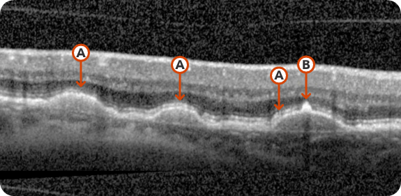
Few medium-sized drusen
- Medium-sized drusen >63 µm and ≤125 µm
- No pigmentary abnormalities
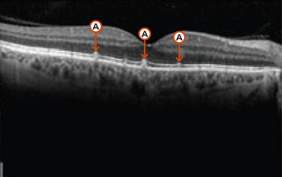
© 2023. This work is licensed under a CC BY 4.0 license. “Correlation between hyperreflective foci and visual function testing in eyes with intermediate age-related macular degeneration”, "Figure 1", Liu TYA, Wang J, Csaky KG. Int J Retina Vitreous.
Drusen grow larger with disease progression
Hyperreflective foci representing RPE migration
- Large drusen >125 µm and/or pigmentary abnormalities
- Can lead to GA, nAMD, or both GA and nAMD
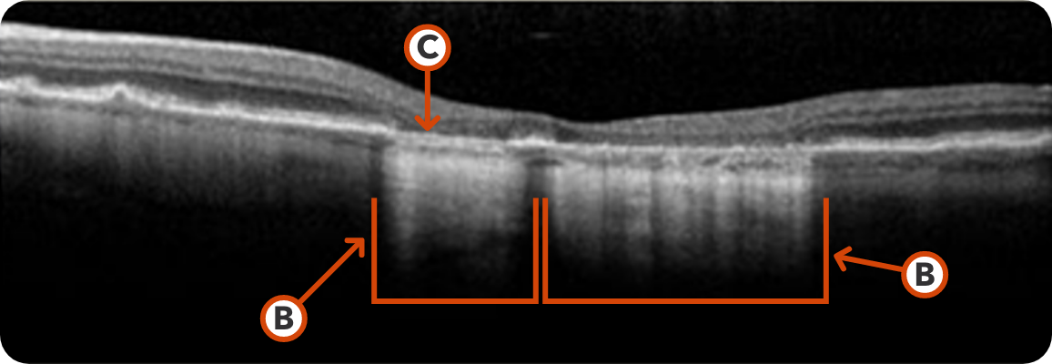
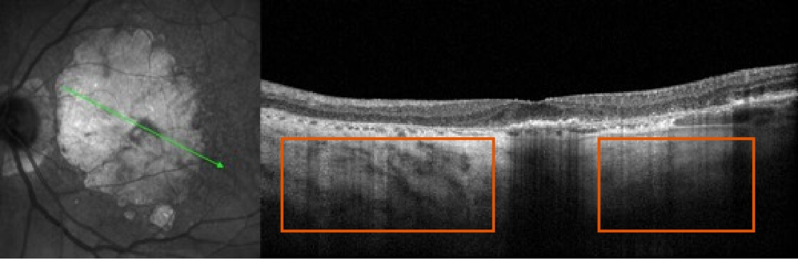
Image courtesy of Mohammad Rafieetary, OD, Charles Retina Institute
Hypertransmission seen in GA
Loss of RPE and photoreceptors
GA4,9-15
- Atrophy of photoreceptors and the loss of RPE can lead to increased reflectivity below Bruch’s membrane and resulting hypertransmission
- Even if visual acuity or BCVA is relatively unchanged, functional vision continues to decline as lesions progress
- Patients with GA can also naturally develop nAMD and vice versa. Up to 29% of patients with GA develop nAMD in about 2 years (N=12,309)*
Images courtesy of Dr. Juan David Arias and Dr. Andrea Hoyos, Colombia.
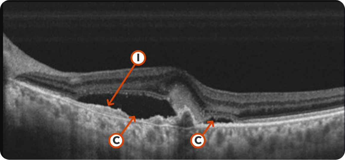
Fluid leakage extending from the choroidal vessels through Bruch’s membrane
Choroidal neovascular membrane
Neovascular AMD (nAMD)10,16
- Distinguished by abnormal blood vessels that may cause fluid or blood to leak into the macula
- Up to 37% of patients with nAMD develop GA in about 2 years (N=91)†‡