SEE THE SIGNS OF GEOGRAPHIC ATROPHY (GA)
GA can destroy so much


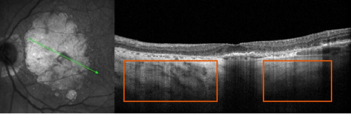
Look for choroidal hypertransmission, a marker of GA in OCT B-scans1
Geographic Atrophy (GA) is an advanced form of age-related macular degeneration (AMD), a leading cause of significant vision loss worldwide. It is defined by atrophic lesions, resulting from loss of photoreceptors, RPE, and underlying choriocapillaris.1-3


It is critical to recognize GA and refer patients in a timely manner, as disease progression is relentless and irreversible1,4-8
Learn how to recognize GA
OCT=optical coherence tomography.